Entamoeba spp
Entamoeba
Updated January 2026
Quick revision
- 4 species – E histolytica, E moshkovskii, E dispar (non-pathogenic), E bangladeshi (uncertain role)
- Worldwide distribution: UK cases are mostly related to travel.
- Reservoir – human; Infective form: cyst; Transmission – faeco-oral, sexual.
- Virulence factor: Surface lectins, Cysteine proteases
- Clinical feature: majority of cases of amoebiasis are asymptomatic (90%). Amoebic dysentery and extraintestinal disease (liver abscess).
- Complications include peritonitis, colonic perforation, and abscess rupture.
- Risk factors: People <50 years old, male, Men having sex with men, Travel to endemic countries, Poor hygiene, institutionalised people, crowding, Alcoholism, immunosuppression, steroids, malnutrition, cancer, syphilis, HIV
- Investigation – stool for OPC, PCR
- Treatment: Treat all patients. Metronidazole is DoC for symptomatic/systemic inf. Diloxanide/paromomycin for asymptomatic (luminal amoebicide). Even after treatment of metronidazole, luminal amoebicide may be needed.
- Abscess – metronidazole (7-10 days) followed by paromomycin/diloxanide
- Drainage needed for Large abscess (>5cm), pt not responding, left lobe abscess, imminent risk of rupture.
Introduction
There are 4 morphologically similar species
- E histolytica causes most diseases.
- E moshkovskii disease is increasing.
- E dispar is non-pathogenic,
- E bangladeshi – role is uncertain [Gilchrist 2014]
Amoebiasis has a worldwide distribution. It is common in resource-poor countries. In the UK, most cases are associated with travel.
Reservoir – human
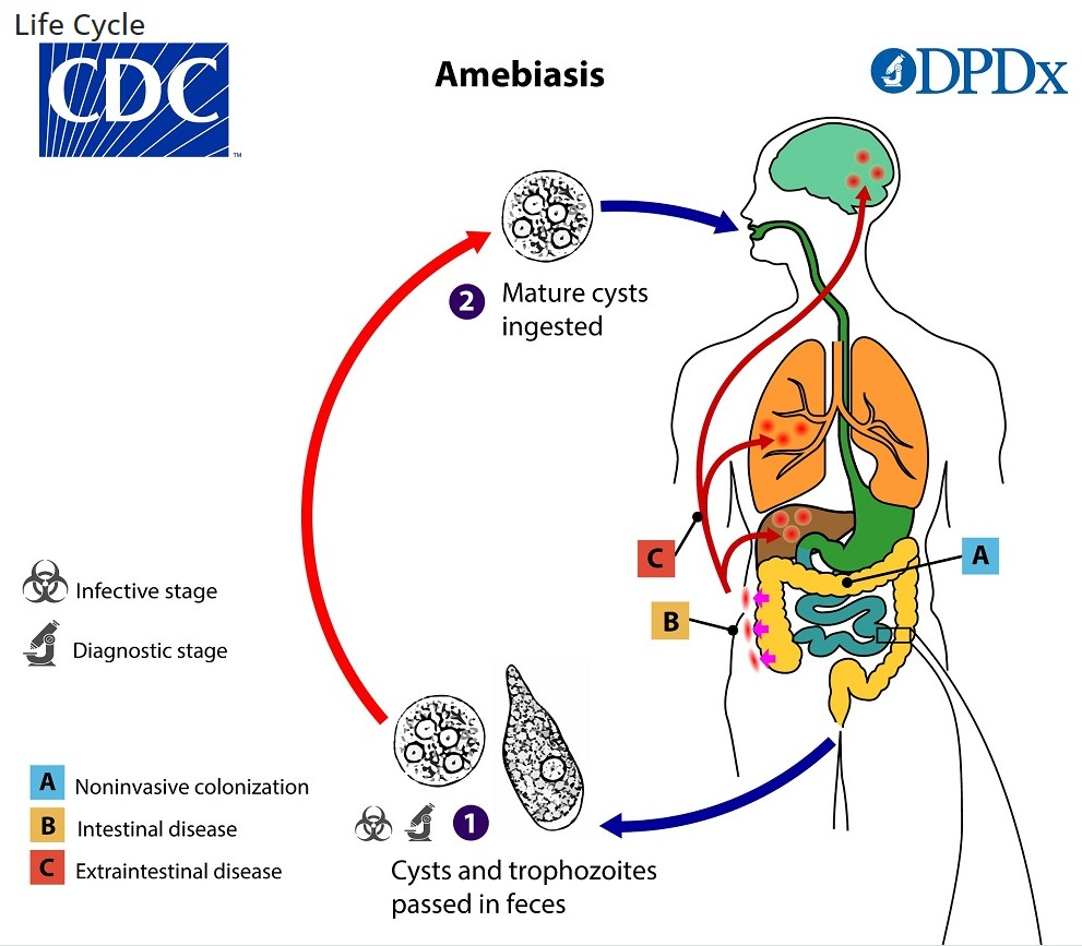
Transmission
- Faeco-oral route; ingestion of contaminated food or water.
- Person to person transmission.
- Sexual transmission – men having sex with men..
Infective form: cyst
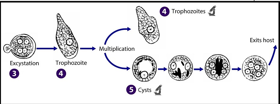
Virulence factors
- Surface lectins: inhibit complement mediated lysis
- Cysteine proteases: degrade Igs
- Host immune modulation: by host cell apoptosis, macrophage migration inhibitory factors
Clinical feature:
- The majority of cases of amoebiasis are asymptomatic (90%) [UKHSA].
It can cause – amoebic dysentery and extraintestinal disease.
Risk factors:
- People <50 years old, male,
- Men having sex with men,
- Travel to endemic countries
- Poor hygiene, institutionalised people, crowding,
- Alcoholism, immunosuppression, steroids, malnutrition, cancer,
- syphilis, HIV
The incubation period
1 – 4 weeks.
| Amoebic dysentery | Fever, diarrhoea. Blood may be present in up to 70% of cases—abdominal pain/tenesmus, and weight loss are most commonly involved. The ascending colon is the most commonly involved part of the bowel. |
| Liver abscess | May present months to years after amoebiasis. The patient may have a fever, right upper quadrant pain, weight loss, and tender hepatomegaly. Alcoholism is a risk factor. Usually, amoebic liver abscesses are large, singular abscesses. |
Complications
Peritonitis, colonic perforation, and abscess rupture.
| ALA is the most common extra-intestinal manifestation of amoebiasis (amoebic dysentery). Entamoeba invades the wall of the large intestine, enters the bloodstream (portal vein), and travels to the liver, where it destroys tissue and forms an abscess. Key Facts & Epidemiology > It most commonly affects men between the ages of 18 and 50 (male-to-female ratio is roughly 10:1) > It is endemic in tropical regions with poor sanitation, including India, Africa, Mexico, and parts of Central/South America > Alcohol consumption, malnutrition, and travel to endemic areas are significant risk factors. Symptoms > Symptoms typically appear within 2 to 4 weeks of infection, though they can sometimes take months to develop > Fever, RUQ pain, hepatomegally (may or may not be present) > Weight loss, nausea, and dry cough. Note that fewer than one-third of patients have active diarrhea or dysentery at the time the abscess is diagnosed Diagnosis > Imaging: Ultrasound or CT scans typically show a single, round, fluid-filled lesion in the right lobe of the liver > Serology (Blood Test): This is the gold standard. Tests for amoebic antibodies are positive in more than 90% of patients. > Aspiration (Needle Test): Rarely required for diagnosis. If performed, the fluid looks like “anchovy paste” (thick, chocolate-brown liquid). It is usually odourless and sterile (no bacteria present). Treatment > Tissue Amoebicide – Metronidazole (750 mg, 3 times daily for 7–10 days) is the standard first-line treatment; alternative – tinidazole > Luminal Agent – After the liver abscess is treated, patients must take a second drug to clear the cysts remaining in the intestine and prevent relapse. Common options include Paromomycin (taken for 7 days) or Diloxanide furoate > Drainage: Aspiration or catheter drainage is not routine. It is reserved for specific high-risk cases: Large abscesses (typically >5–10 cm). Abscesses in the left lobe Failure to respond to antibiotics after 3–5 days. Imminent risk of rupture Differential > Pyogenic Liver Abscess (Streptococcus constellatus/anginosus): a scenario of a liver abscess associated with colonic carcinoma or underlying malignancy. The organism answer in these cases is usually Streptococcus constellatus (part of the Strep. milleri group) rather than Entamoeba |
Diagnosis
Tests
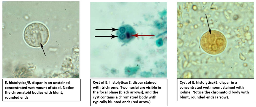
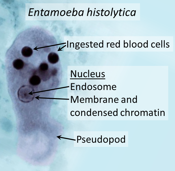
Available tests are – stool microscopy, stool antigen detection, stool polymerase chain reaction (PCR), serology, and colonoscopy with histologic examination.
PCR is the method of choice for diagnosis of E histolytica symptomatic and asymptomatic cases [UKHSA].
Stool microscopy is commonly used in many labs.
Management
- Treat all patients with Entamoeba histolytica infection, whether or not symptoms are present [WHO].
- Metronidazole is used as the drug of choice for acute invasive amoebic dysentery and amoebic abscess.
- Diloxanide furoate is the drug of choice for asymptomatic patients with E. histolytica cysts in the faeces.
- A 10-day course of diloxanide furoate should be given after metronidazole or tinidazole treatment to destroy any amoebae in the gut. [BNF]
- Duration for metronidazole – 5 days in intestinal infection & for 5–10 days in extra-intestinal infection (followed by diloxanide, as above).
Large abscess (>5cm diameter) may need drainage.
Tissue amoebicide.- metronidazole, tinidazole, ornidazole, secnidazole, and nitazoxanide.
Intestinal/luminal amoebicides – paromomycin, iodoquinol, and diloxanide furoate.
- Universal enteric precautions and exclusion for 48 hours after the resolution of diarrhoeal symptoms should be followed for all cases with diarrhoea
- No additional formal exclusion periods or microbiological clearance for public health reasons for confirmed cases of E. histolytica infection are required.
- Cases should undergo screening one week after the completion of treatment for clinical reasons to confirm treatment success.
- Household, co-traveller and sexual contacts of a confirmed case should be tested for asymptomatic E. histolytica infection to facilitate early treatment.
Ref:
https://www.gov.uk/government/publications/amoebiasis-public-health-operational-guidelines
| Recent developments |
| Trogocytosis and immune evasion: Trogocytosis is a phenomenon by which a cell bite of part of another cell (as opposed to phagocytosis where the cell eat another cell/pathogen). This trogocytosis is seen in lymphocytes when the nibble part of antigen presenting cells. Recently this phenomenon has been described in E histolytica. Amoebae kill human cells by performing trogocytosis. After performing trogocytosis, amoebae display human proteins (CD46 or CD55) on their own surface and become resistant to lysis by human complement system. https://www.biorxiv.org/content/10.1101/2024.10.04.616735v1When E. histolytica is exposed to enteropathogenic E. coli (gut bacteria), it exhibits “superdiffusive” movement. The bacteria physically interact with the amoeba, causing it to move faster and with greater directionality, which likely increases tissue damage and invasiveness. PLOS may 2025 |